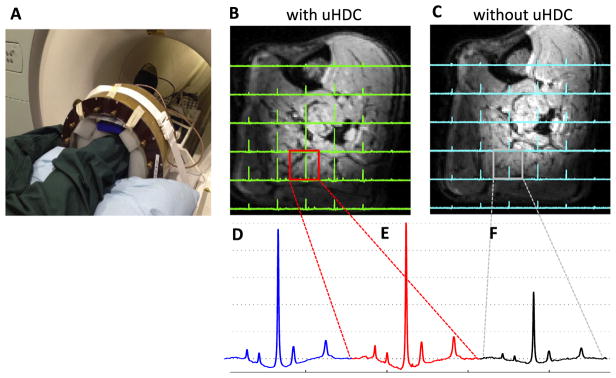
Fig. 3.
(A) Photograph of experimental setup for the in vivo 31P MRS study of the human calf muscle with the placement of four uHDC blocks around the subject’s leg. In vivo 31P CSI profiles with the uHDC blocks at a RF pulse reference voltage of 63 V (B), and without the blocks at a reference voltage of 141 V (C). (D) Comparison of in vivo 31P spectra acquired using the small 31P surface coil at a reference voltage of 60 V, and the 31P volume coil with (E) and without the uHDC blocks (F).

