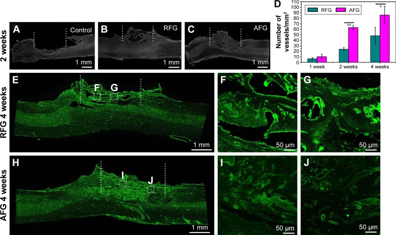
Figure 2.
Vascularization in the dorsal hemisected site. (A–C) Representative sagittal sections of control, RFG, and AFG groups stained by DAPI show the tissue regeneration at 2 weeks post-surgery, and the lesion site region is labeled by dashed line. (D) Quantification of RECA-1 positive vessels at the middle site of injury at 1, 2, and 4 weeks after spinal cord injury. *p < 0.05, **p < 0.01 for the comparison of RFG and AFG. Four individual rats per group were used for statistical analysis (n = 4), and five confocal images per rats were used for the vessel counts. Immunofluorescence staining images of the T8–T10 sagittal sections in RFG (E–G) and AFG (H–J) groups labeled by RECA-1 (green).
Abbreviations: AFG, aligned fibrin hydrogel; DAPI, 4′,6-diamidino-2-phenylindole; RFG, random fibrin hydrogel.

