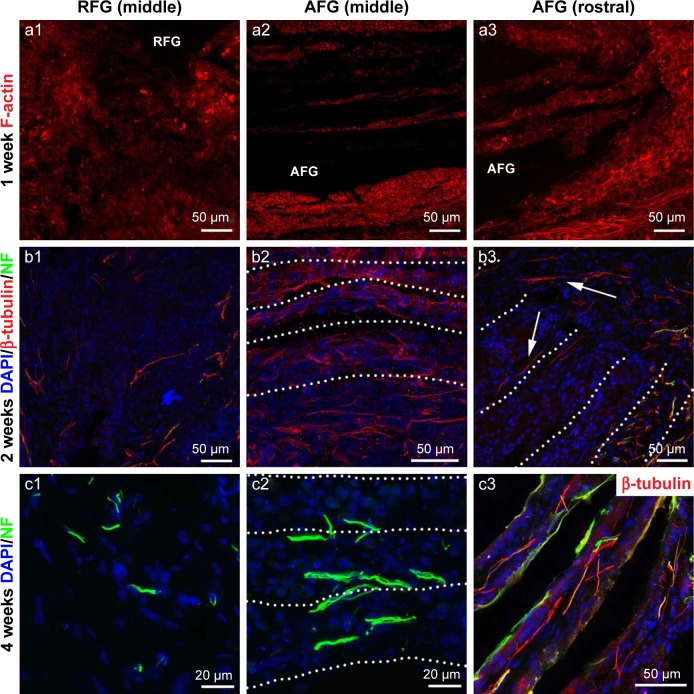
Figure 3.
Aligned tissue cables formed by the endogenous cell invasion promote extensive axonal regeneration. (a1–a3) F-actin immunofluorescence staining at 1 week after surgery. The representative immunofluorescence staining images of β-tubulin III (red), NF (green), and DAPI (blue) at 2 weeks (b1–b3) and 4 weeks (c1–c3) after surgery. The dashed lines indicate the long axis of the aligned AFG and cell cables. The arrows in b3 show the cells (neurons) labeled by β-tubulin III migrate into the regrowth tissue.
Abbreviations: AFG, aligned fibrin hydrogel; DAPI, 4′,6-diamidino-2-phenylindole; NF, neurofilament; RFG, random fibrin hydrogel.

