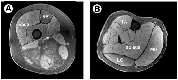
Fig 1.
T1-weighted fat-suppressed transaxial images of the thigh (A) and lower leg (B) obtained from an 11-year-old boy with DMD. The rectus femoris and vasti muscles are outlined on the thigh, and the TA, soleus, MG, and LG are outlined on the lower leg. Note the presence of fatty infiltration to a greater extent in the thigh musculature than in the calf. Abbreviations: LG, lateral gastrocnemius; MG, medial gastrocnemius; RF, rectus femoris; TA, tibialis anterior.

