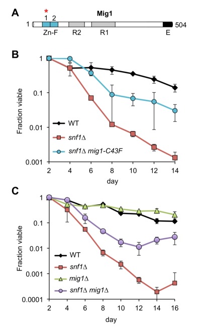
Figure 3. FIGURE 3: Mutations in MIG1 partially restore CLS to a snf1∆ strain.

(A) Diagram of Mig1 protein domain organization showing the two zinc fingers (Zn-F), two regulatory domains (R1 and R2) and an effector domain (E) 36. Red asterisk indicates the zinc finger mutated (C43F) in suppressor candidate JS1430.
(B) Quantitative CLS assay showing short CLS of snf1∆ (JS1394) compared to WT (JS1256). The snf1∆ mig1-C43F mutant (JS1430) extends CLS compared to snf1∆.
(C) Quantitative CLS assay showing similar partial CLS extension by deleting MIG1 in the snf1∆ background (JS1446). Deletion of MIG1 had little CLS effect on its own (JS1442). CLS assays were performed in SC 2% glucose conditions. Error bars indicate standard deviation.
