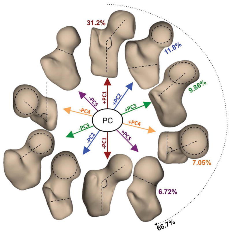
Figure 1.

Description of the first five 3D bone shape modes extracted from a dataset of 80 MRI exams. Each principal component (mode) is visualized at mean +- 3standard deviation. PC1 (Mode 1): neck-shaft angle in coronal plane (coxa valga/coxa vara); PC2 (Mode 2): ratio between the femoral head radius and the shaft thickness; PC3 (Mode 3): medial–lateral length of the femoral neck; PC4 (Mode 4): neck shaft angle in the axial plane. PC5 (Mode 5): thickness of femoral neck and sphericity of the femoral head (pistol grip deformity)
