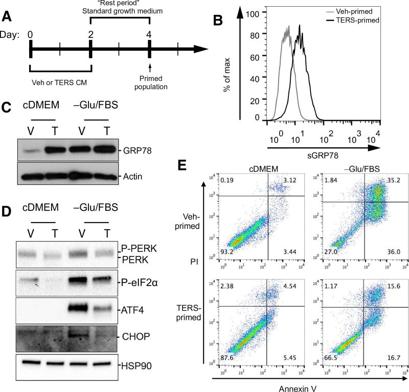
Fig. 2. TERS-primed cancer cells display a unique UPR and are protected against nutrient deprivation.
(A) Treatment design of TERS priming: 2-day culture in Veh CM or TERS CM followed by 2-day rest period. Cells were then challenged and analyzed as indicated. (B) Flow cytometry analysis for surface abundance of GRP78 in vehicle- or TERS-primed PC3 cells grown in standard growth medium. (C) Western blot analysis of GRP78 in vehicle (V)– or TERS (T)–primed PC3 cells after 48-hour culture in standard growth medium (cDMEM) or in nutrient-deprived condition [-Glu/FBS (fetal bovine serum)]. (D) Western blot analysis of proteins of the PERK pathway in vehicle- or TERS-primed PC3 cells after 48-hour culture in cDMEM or in -Glu/FBS. (E) Apoptosis analysis by flow cytometry detection of annexin V in vehicle- or TERS-primed PC3 cells after 48-hour culture in cDMEM or in -Glu/FBS (each plot represents at least 10,000 events per condition). Data in (D) are representative of two experiments; data in (B), (C), and (E) are from three or more independent experiments.

