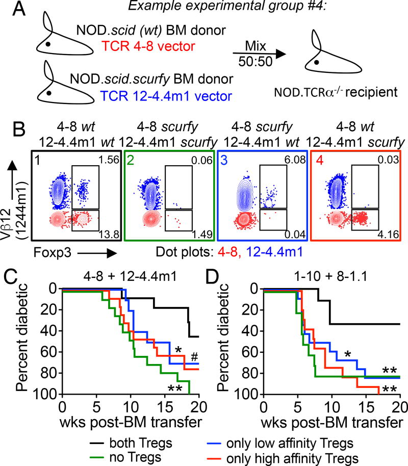
Figure 1.
Both high and low affinity Tregs contribute to protection during autoimmunity. (A) An example of a two-TCR retrogenic chimera experimental group where Foxp3+ Treg development is limited to 4–8 TCR expressing T cells. (B) Representative flow plots of Foxp3+ Tregs from the spleen of 4–8/12-4.4m1 two-TCR Rg BM chimeras. Analysis is gated on CD4+CD3+ cells, Vβ12+ (blue) are 12-4.4m1, and Vβ12– (red) are 4–8 T cells. Mice were analyzed 5.3 weeks post-bone marrow transfer. (C and D) Diabetes incidence for two-TCR Rg chimeras expressing either 4–8 and 12-4.4m1 (C) or 1–10 and 8-1.1 TCRs (D). Mice were monitored for spontaneous diabetes development for 20 weeks (n=10–13 mice per group, #p=0.057). Data are pooled from six (C) and nine (D) independent experiments.

