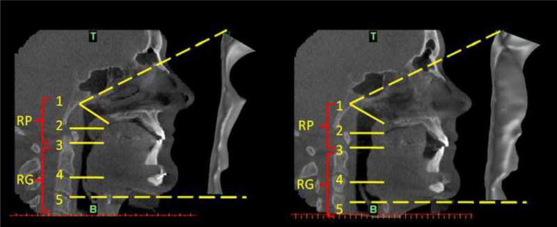
Figure 1.

SDB Patient #5: Pre-treatment and post-treatment CT sagittal views of the upper respiratory tract together with side views of the reconstructed airway models at pre and post-treatment conditions: pre-treatment CT image and pre-treatment reconstructed airway volume (left); post-treatment CT image and post-treatment reconstructed airway volume (right). The anatomical locations marked on the CT images with RP and RG refer to the retro-palatal and retro-glossal regions, while zones 1, 2, 3, 4, and 5 refer to: 1- nasal choanae level (T is top face of the computational domain); 2 - the minimum cross-sectional area (CSA) in the RP region at pre-treatment condition; 3 - tip of uvula; 4 - tip of epiglottis; 5 - base of epiglottis (B is bottom face of the computational domain).
