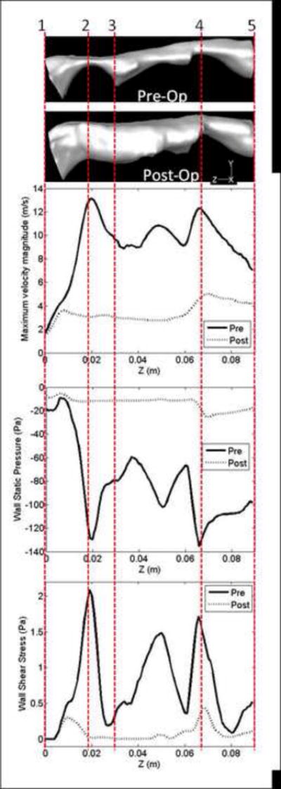
Figure 4.

SDB Patient #5: Comparison of flow variables along the pharyngeal airway during inspiration at pre and post-treatment conditions: maximum velocity magnitude (m/sec) along the airway; wall static pressure (Pa) distribution along the anterior airway wall in the mid-sagittal plane of the airway, and wall shear stress (Pa) distribution along the anterior airway wall in the mid-sagittal plane of the airway. Data obtained with steady-state RANS (k-ω SST model). The zones 1 to 5 (see also Figure 1) are marked on the horizontal side views of the pre and post-treatment pharyngeal airway models.
