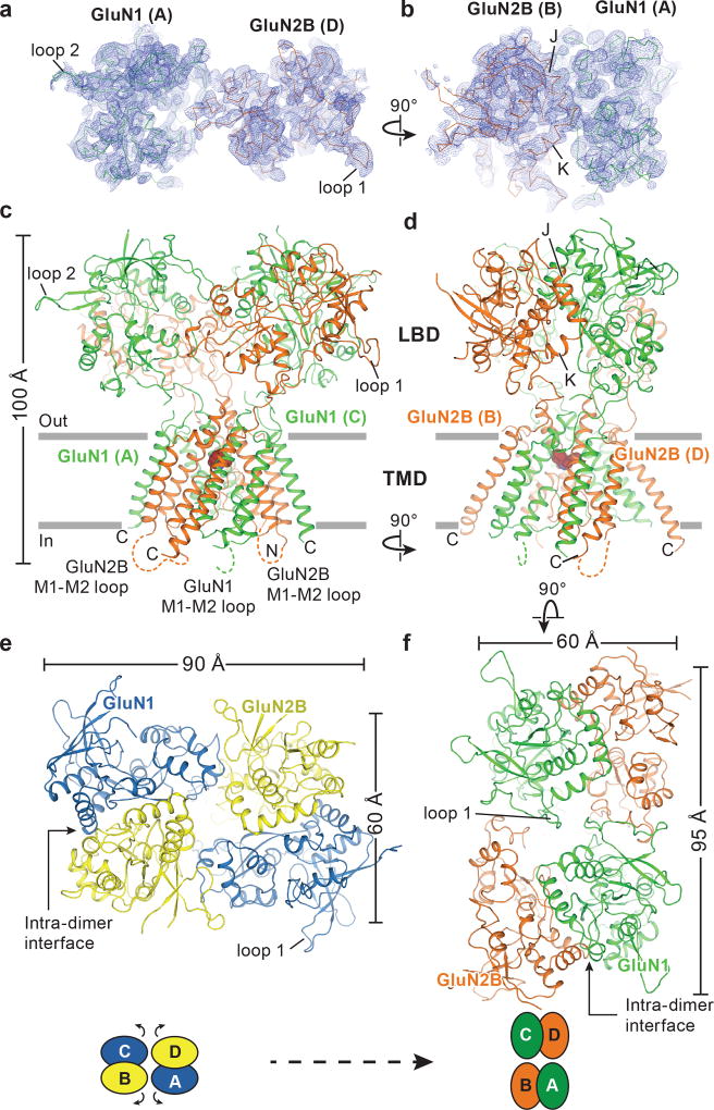
Figure 1. Architecture of the GluN1/GluN2B ∆ATD NMDA receptor.
a-b, Composite omit maps (blue mesh) of GluN1 (green ribbon) and GluN2B (red ribbon) LBDs contoured at 1.0 σ, showing the inter-dimer (a) and intra-dimer interfaces (b). c, d, Side views of the ∆ATD receptor with GluN1 (green) and GluN2B (orange) subunits. e-f, Top-down views of the ∆2 receptor (e) and the ∆ATD receptor (f) from the extracellular side of the membrane.

