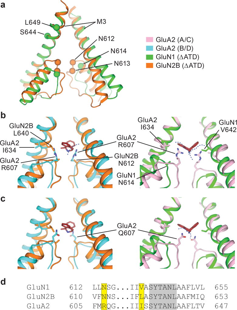
Figure 3. Steric clashes block MK-801 binding at AMPA receptors.
a, Superposition of the M2, pore loop and M3 elements of the GluN1 (green) and GluN2B (orange) subunits from the ∆ATD receptor crystal structure. The α carbons of key asparagine residues are shown as spheres. b-c, Superposition of elements of the ∆ATD receptor from panel a with the equivalent elements of the GluA2 AMPA receptor (PDB code: 5VOT) with R607 (b) or Q607 (c). The NMDA receptor GluN2B subunits (orange) and GluN1 subunits (green) are superposed on the equivalent regions of the GluA2 AMPA receptor B/D subunits (cyan) or the A/C subunits (pink), respectively. All superpositions are based on the Cα atoms of the conserved ‘SYTANL’ region. Dashed lines show likely steric clashes. d, Sequence alignment of the channel region between the NMDA receptor and AMPA receptor subunits. The residues involved in MK-801 binding and the corresponding GluA2 residues are highlighted in yellow. Residues of the ‘SYTANL’ motif are highlighted in gray.

