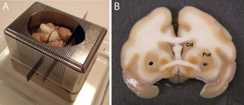
Figure 5.
Brain processing post-necropsy. (A) Following removal at necropsy, the brain is placed in to a matrix and sectioned into slabs of varying thickness using tissue slicer blades. (B) The slabs are placed into petri dishes containing sterile saline and biopsy punches are taken from key areas to be used for molecular and biochemical studies. Brain slabs are then post-fixed in 4% paraformaldehyde for 48 hours and cryoprotected in 30% sucrose prior to immunohistochemical staining.

