
Fig. 5.
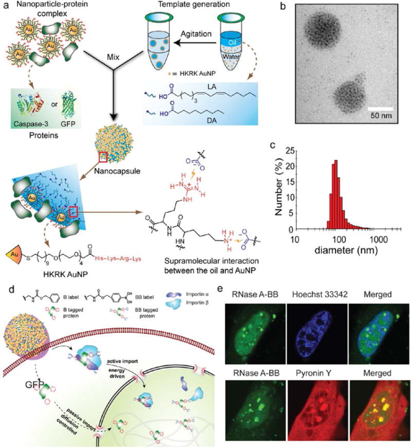
Design, preparation and delivery mechanism of NPSCs. a) Schematic showing the preparation of the protein-NPSC complex containing caspase-3 or GFP. b) TEM image of dried GFP-NPSC. c) DLS histogram of GFP-NPSCs indicating an average diameter of ∼130nm (Adapted with permission from Ref. 19. Copyright (2013) American Chemical Society). d) Schematic diagram showing the delivery of proteins tagged with benzyl boronate (BB) to the cytosol followed by translocation to the nucleus using active transport with boronate ligands and through passive diffusion using nonboronate analogues. e) Colocalization of RNase A-BB with Hoechst 33342, a DNA staining dye (top) and with Pyronin Y, a dsRNA staining dye (bottom) (Adapted with permission from Ref. 39 Copyright (2017) American Chemical Society).