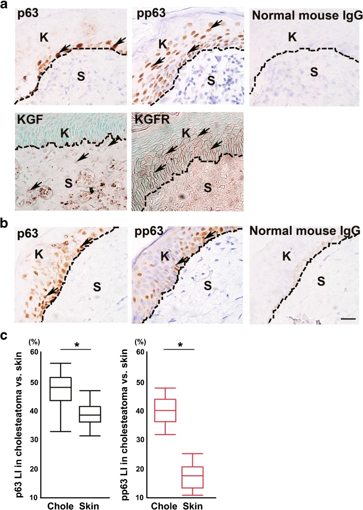
Fig. 1.
Immunohistochemical analysis of KGF, KGFR, p63, and pp63 in human specimens. a p63-positive cells were detected mainly in the basal layer; pp63-positive cells were detected in the basal and upper layers of the cholesteatoma matrix (upper panels). KGF expression was detected in some stromal cells, and KGFR-positive cells were detected in the basal and upper layers of the cholesteatoma matrix (lower panels). Normal mouse IgG was used instead of first antibody as a negative control. b p63-positive cells and pp63-positive cells were detected in the basal and upper layers and the expression level was slightly weak in the control. Normal mouse IgG was used instead of first antibody as a negative control. Arrows indicate positive cells. Dashed lines indicate the basement membrane. K indicates keratinizing squamous epithelium. S indicates the subepithelial region. c Box plot showing the labeling index (LI) of p63 (black boxes) in cholesteatoma (n = 29) vs. skin (n = 22) and pp63 (red boxes) in cholesteatoma (n = 29) vs. skin (n = 21). (p63 LI: *p < 0.0001 as determined by a one-way ANOVA F(1, 49) = 36.22 with a Tukey’s post hoc test, pp63 LI: *p < 0.0001 as determined by a one-way ANOVA F(1, 48) =294.20)

