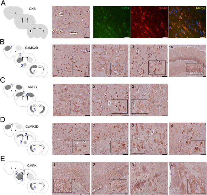
Figure 4.
Histological localization of protein candidates in the ischemic brain. (A) CKB, (B) CaMK2B, (C) AREG, (D) CaMK2D and (E) CMPK histological examination in three established cortical depths of the ischemic brain (see Supplemental Figure S1). In the schematic representation of brain, right hemisphere corresponds to IP and left hemisphere corresponds to CL. Dark grey regions indicate higher abundance of positive staining than pale gray zones. CKB (green) was co-localized with GFAP marker in the red channel and nuclear detection was conducted with DAPI in the blue channel. Scale bar = 50 µm.

