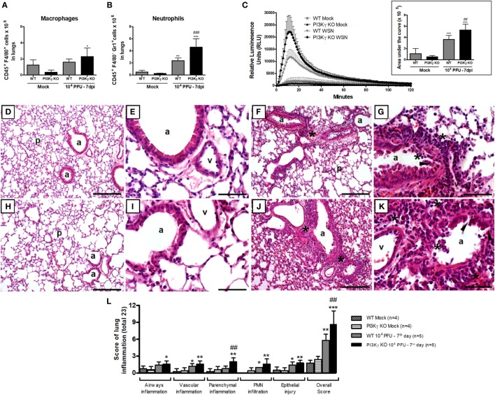
Figure 8.
Neutrophil recruitment and activation and lung damage during influenza A virus infection are affected by phosphatidyl inositol 3 kinase-gamma (PI3Kγ). WT and PI3Kγ knockout (KO) mice infected with 104 plaque-forming units (PFU) of WSN or receiving phosphate-buffered saline (Mock) were killed at day 7 after infection (n = 4–7 mice per group). (A) Macrophage (CD45+F4/80+) and (B) neutrophils (CD45+F4/80−GR1+) numbers in lungs assessed by flow cytometry. (C) Reactive oxygen species assessed in lung leukocytes in presence of luminol with or without zymosan for 120 min. Relative luminescence units mean the value with zymosan minus the respective value without zymosan. Areas under the curve were compared using mean ± SEM and one-way ANOVA, Newman–Keuls test (D–K). Representative slides of hematoxylin and eosin stained lung sections from WT (D,E) and PI3Kγ KO mock (H,I), WT (F,G), and PI3Kγ KO-infected mice (J,K) are shown in 100× [(D,H,F,J), bars = 200 μm] and 400× [(E,I,G,K), bars = 50 μm] magnification. “p” Indicates lung parenchyma, “v” vessels, and “a” airways; asterisks foci of inflammatory infiltrates; and arrowheads areas of epithelial injury. (L) Histopathological score (total 23 points) assessed airway, vascular, parenchymal inflammation, neutrophilic infiltration, and epithelial injury. Data are presented as mean ± SD. *p < 0.05, **p < 0.01, and ***p < 0.001, compared with each Mock group; ##p < 0.01 and ###p < 0.001, compared with WT-infected mice (one-way ANOVA, Newman–Keuls).

