Figure 3.
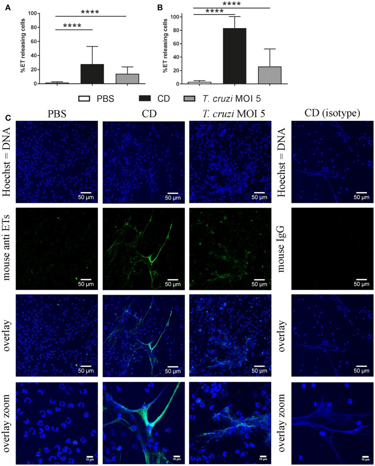
T. cruzi induces ETs in didelphine granulocytes in vitro. Formation of ETs after infection of purified didelphine granulocytes with T. cruzi at MOI of 5 after 60 min (A) and 180 min (B) was analyzed. PBS was used as negative control and the cholesterol-depleting cyclodextrine (CD) as positive ET inducer. After infection ETs were visualized by immunofluorescence microscopy (ETs [DNA/histone-1-complexes] = green, Hoechst [DNA] = blue). Per sample at least 6 pictures were taken at predefined positions, and the number of granulocytes and the ET-releasing granulocytes were determined. (C) Representative pictures after 60 min of stimulation are shown. The isotype control was negative for ETs. Graphs in A and B show the mean ± SD of 4 independent experiments with 24 analyzed pictures. Statistical differences were tested by unpaired one-tailed Students t-Test (****p < 0.0001).

