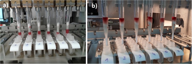
FIGURE 5.

Filter wand’s status (a) at the end of the 2-min vacuum aspiration in the lyzed blood cells and (b) at the end of wash 2 step showing that all of the aspirated liquids are contained in the filter wand body and blood components are moved far away from the filtration membrane to limit the pollution of MALDI-TOF MS slide with hemoglobin during the spotting step.
