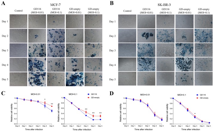
Figure 3.
Cytotoxicity of GD116 and GD-empty in MCF-7 and SK-BR-3 cells in vitro. Monolayers of cells were mock-infected or infected with viruses at multiplicities of infection of 0.01 and 0.1 and further incubated at 37°C for 5 days. (A and B) X-gal staining of MCF-7 and SK-BR-3 cells. Infected cells expressing LacZ were stained positive (blue) with X-gal. Original magnification, ×100. (C and D) Cell viability was assessed using an MTT assay from days 1 to 5 following infection. The relative cell viability was normalised to that of the mock-infected control. Statistical comparisons (independent t-test) were between two viruses at the indicated times. *P<0.05 and **P<0.01.

