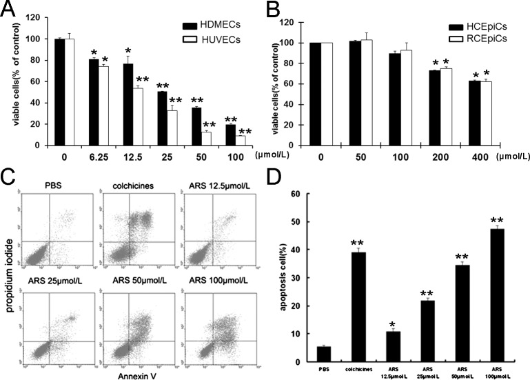
Figure 2.
Effects of ARS on HUVECs proliferation and apoptosis. (A) HDMECs and HUVECs were treated with ARS at indicated concentrations for 48 hours. The viable cells were quantified by MTT assay. (B) RCEpiCs and HCEpiCs were treated with ARS at different concentrations for 48 hours. The viable cells were quantified by MTT. (C) After incubation with ARS for 24 hours, apoptotic HUVECs were stained with AnnexinV and PI, and analyzed by flow cytometry. (D) Quantitative analysis of apoptosis in endothelial cell induced by ARS. n = 3, * and ** denote P < 0.05 and P < 0.01, respectively.

