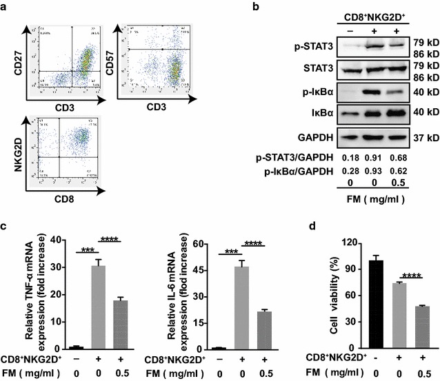
Fig. 2.

FM mitigates CD8+NKG2D+ cell-activated inflammatory signaling and enhances the oncolysis of CD8+NKG2D+ cells. a CD8+NKG2D+ cells were generated as described in methods. Cells were identified by flow cytometry with anti-CD3, anti-CD8, anti-NKG2D, anti-CD27 and anti-CD57 antibodies. b–d HCCLM3 cells stably expressing luciferase (HCCLM3-luciferase) (target cells, T) were pre-treated with FM at a non-toxic dose of 0.5 mg/ml for 12 h or left untreated, then medium was discarded. CD8+NKG2D+ cells (effector cells, E) were added at a ratio of 5:1 (E: T) in fresh medium for 12 h. Then, cells were harvested for western blot, qRT-PCR analysis, or luciferase activity measurement. b The protein level of IκBα, phosphorylated IκBα, STAT3 and phosphorylated STAT3, c mRNA level of TNF-α (left panel) and IL-6 (right panel), and d luciferase activity reflecting the cell viability are shown. GAPDH was used as loading control. Similar results were obtained in two independent experiments. Mean ± SD of each group are shown. ***p < 0.001, ****p < 0.0001
