Fig. 1.

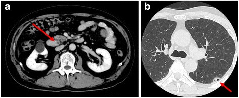
Computed tomography findings. a Abdominal computed tomography showing thickening of the distal bile duct (arrow). b Chest computed tomography showing an air-space consolidation in the upper lobe of left lung (arrow)

Computed tomography findings. a Abdominal computed tomography showing thickening of the distal bile duct (arrow). b Chest computed tomography showing an air-space consolidation in the upper lobe of left lung (arrow)