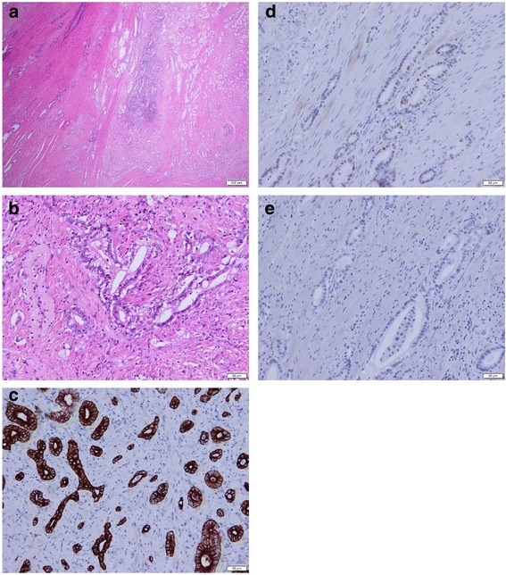
Fig. 2.

Hematoxylin and eosin (H&E) staining and immunohistochemistry of the resected specimen after pancreaticoduodenectomy. H&E stains show well-differentiated to moderately differentiated adenocarcinoma (a, original magnification × 10; b, original magnification × 200). Immunohistochemical stains show that the tumor cells were positive for cytokeratin 7 (c, original magnification × 200) and CDX-2 (d, original magnification × 200) and negative for cytokeratin 20 (e, original magnification × 200)
