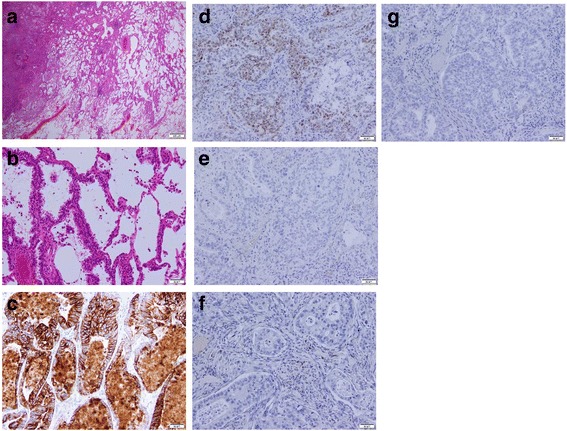
Fig. 5.

The resected lung specimen resembled the previous distal cholangiocarcinoma. Histopathological examination (H&E staining) of the lung tumor showed tumor cells forming irregular tubular structures with a lepidic pattern (a, original magnification × 10; b, original magnification × 200). Immunohistochemically, the tumor cells were positive for cytokeratin 7 (c, original magnification × 200) and CDX-2 (d, original magnification × 200) and negative for thyroid transcription factor 1 (e, original magnification × 200), napsin A (f, original magnification × 200), and cytokeratin 20 (g, original magnification × 200)
