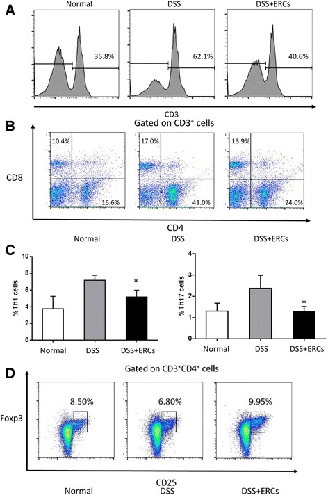
Fig. 3.

The regulatory effects of endometrial regenerative cell (ERC) treatment on T lymphocytes. The spleen was dissected and made into a single-cell suspension. Cells were stained with fluorescently labeled CD3, CD4, CD8, CD25, IFN-γ, IL-17, and Foxp3, and detected by flow cytometry. The proportion of a CD3+ T cells in lymphocytes, b CD4+ and CD8+ T cells in CD3+ T cells, c IFN-γ+ and IL-17+ T cells in lymphocytes, and d CD25+Foxp3+ Tregs in CD3+CD4+ T cells was detected by flow cytometry. Graphs represent mean ± SEM of triplicate experiments. P value was determined by one-way ANOVA. *P < 0.05. DSS, dextran sodium sulfate; Th, T helper
