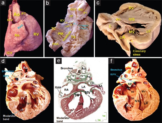
Figure 2.

(a) Lateral view of the RA and its appendage, a roughly triangular-shaped offshoot whose apex generally points upward, overlapping the aortic root. (b) The RA and ventricle have been opened like a book to show the different components of the RA, such as the terminal crest and the origin of the pectinate muscles, the vestibule next to the tricuspid valve, the triangle of Koch and the orifice of the coronary sinus. (c) Sagittal section of a 28-week fetal heart showing the coronary sinus which extends through the left atrioventricular groove and is separated from the walls of the LA. (d-f) Frontal sections of a 23-week heart, where image e is a histologic stain using trichromic Masson stain corresponding to image d. Note the different morphology of the right and left atrial appendages and distinguish between the smooth-walled LA and the pectinated RA. Note in image f, the superior interatrial fold between the right pulmonary veins to the LA, and the caval veins to the RA. Ao: Aorta, IVS: Interventricular septum, CS: Coronary sinus, CSO: Coronary sinus orifice, E: Esophagus, EV: Eustachian valve, IVS: Interventricular septum, LA: Left atrium, LAA: Left atrial appendage, LAAO: Left atrial appendage orifice, LV: Left ventricle, LSPV: Left superior pulmonary vein, MV: Mitral valve, OF: Oval fossa, PA: Pulmonary artery, PM: Pectinate muscles, PT: Pulmonary trunk, RA: Right atrium, RAA: Right atrial appendage, RIPV: Right inferior pulmonary vein, RV: Right ventricle, SCV: Superior caval vein, SIF: Superior interatrial fold, TC: Terminal crest, TV: Tricuspid valve
