
Figure 4.
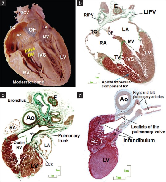
(a and b) Frontal sections of a 26-week heart, where image b is a histologic stain using trichromic Masson stain corresponding to image a. Two of the three components of the RV are revealed: The inlet (tricuspid valve), and the apical trabecular component. Note an important muscular strand, the moderator band, to the anterior papillary muscle and the septal surface reinforced by the septomarginal trabeculation (c and d). Histological sections of the outlet of the RV stained with Masson trichrome of two fetus of 22 (c) and 27 weeks/day) showing the free-standing infundibular sleeve and the attachment of the pulmonary leaflets. Note the anatomic relation of the RV outflow tract with the aortic outflow. Ao: Aorta, E: Esophagus, IVS: Interventricular septum, LA: Left atrium, LCx: Left circumflex artery, LV: Left ventricle, LIPV: Left inferior pulmonary vein, MV: Mitral valve, OF: Oval fossa, RA: Right atrium, RIPV: Right inferior pulmonary vein, RV: Right ventricle, SCV: Superior caval vein, TC: Terminal crest, TV: Tricuspid valve