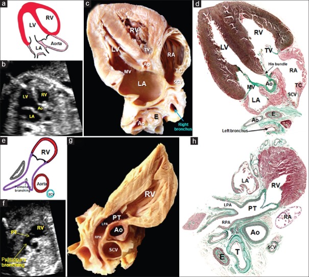
Figure 7.
(a-d) A composite of drawing (a), echocardiogram (b), dissection (c), and histological section (d) illustrating the findings in the third plane: the aortic outflow tract, also called the echocardiographic cut “five-chamber view” showing the aorta leaving the LV in a central position, the subaortic membranous septum, and the area of mitroaortic continuity. (e-h) A composite of drawing (a), echocardiogram (f), dissection (g), and histological section (h) illustrating the fourth plane: the short axis of the right ventricular outflow tract. In this section, T is possible to discern the features of ventriculoarterial concordance, as well as the support of the pulmonary valve and the branching of the PT into the right and left pulmonary arteries. Ao: Aorta, E: Esophagus, LA: Left atrium, LPA: Left pulmonary artery, LV: Left ventricle, MV: Mitral valve, PT: Pulmonary trunk, RA: Right atrium, RPA: Right pulmonary artery, RV: Right ventricle, SCV: Superior caval vein, TC: Terminal crest, TV: Tricuspid valve

