
Figure 1.
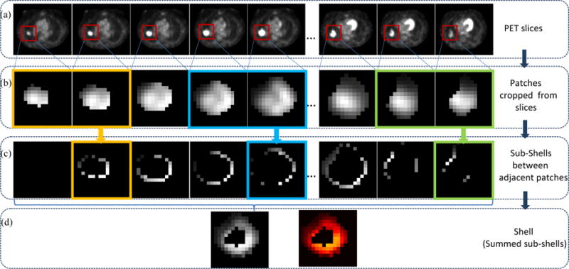
Shell feature extraction workflow. (a) Series of axial PET slices of one patient, (b) Series of patches (red windows in (a)) including tumors are cropped from each slice in (a), (c) Series of sub-shells derived from adjacent two patches in (b), (d) Shell feature, with grayscale image left and Heatmap image right.