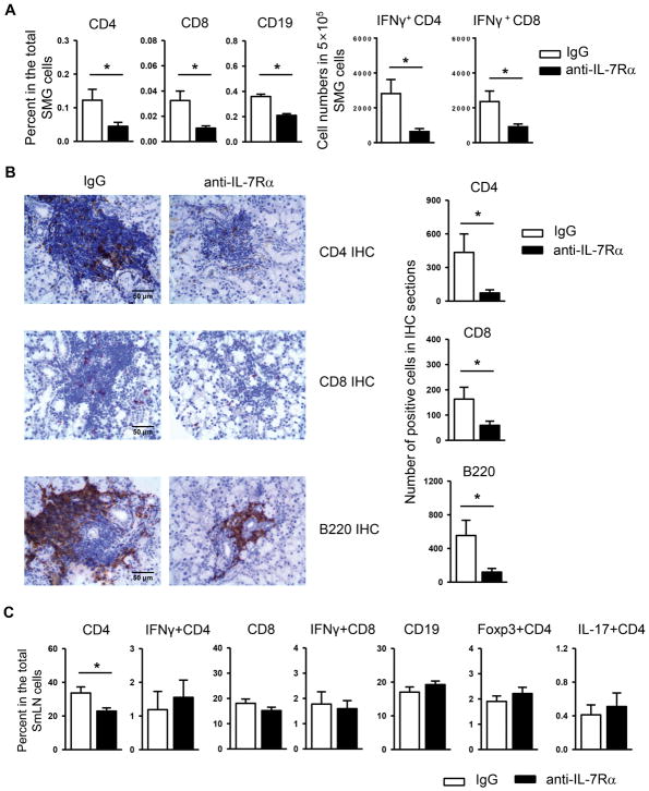
Figure 2. Blockade of IL-7Rα causes a reduce of T and B cell accumulation in the SMGs of NOD mice.
Anti-IL-7Rα antibody or control IgG was i.p.-administered to 10-week-old female NOD mice, 3 times weekly for 3 weeks. (A) Flow cytometric analysis of the percentage of lymphocyte populations among total SMG cells, left panels, and the number of IFN-γ+CD4 and IFN-γ+CD8 T cells among the 5×10⁵ total SMG cells analyzed, right panels. (B) Immunohistochemical staining of SMG sections for CD4, CD8 and B220 (scale bar = 50μm). Bar graph shows the quantification of the number of positively-stained cells. (C) Flow cytometric analysis of the percentage of lymphocyte populations among total submandibular lymph node (smLN) cells. Data are the average of analyses of 7 mice each group.

