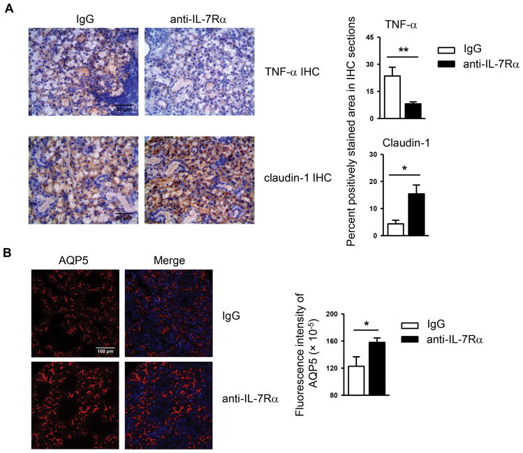
Figure 4. Blockade of IL-7Rα reduces TNF-α levels and increases claudin-1 and AQP5 amounts in the SMGs.
Anti-IL-7Rα antibody or control IgG was i.p.-administered to 10-week-old female NOD mice 3 times weekly for 3 weeks. (A) Immunohistochemical staining of TNF-α and claudin-1 protein in SMG sections (scale bar = 50μm). Bar graph shows the percentage of positively stained areas in the sections. (B) Immunofluorescence staining of AQP5 protein in SMG sections (scale bar = 100μm). Bar graph shows the fluorescence intensity of AQP5 staining. Data are representative or the average of analyses of 7 mice each group.

