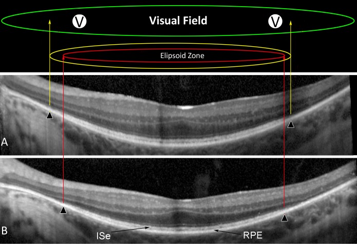
Figure 2.
Ellipsoid zone anatomic and EZ transitional visual field sensitivity. Identified by semiautomated segmentation of retinal frequency-domain OCT from participant G101 (segmentation lines not shown) is the dense layer of outer photoreceptor segments termed the EZ which lies between the hyperreflective ISe and the hyperreflective RPE band. The anatomic EZ width was defined by the nasal and temporal points where the outer segment layer has declined to zero (▴) at trial year 2 (A) and year 4 (B). By superimposing the visual fields over the segmented scans, two nasal visual field locations just inside the EZ edge were selected for each patient for evaluation of EZ transitional visual field sensitivity (V).

