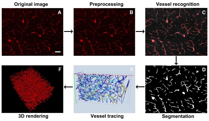
Figure 1.
Processing procedures of clarified sample image. A) Original image obtained from the clarified sample. Scale bar=100 μm. B) Image preprocessing to uniform background intensity of the image. C) Vessel recognition by 3D Canny edge detection and morphology operation. D) Image segmentation and binarization. E) Vessel tracing using Vaa3D software. F) Visualization of 3D rendering images was performed by Vaa3D.

