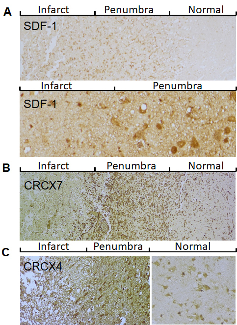
Figure 1.

Expression pattern of SDF-1/CXCR4/CXCR7 in post-stroke human brain. A) Representative images show that SDF-1 expression in cerebral cortex of infarcted brain. Top panel: low magnification; Bottom panel: high magnification. B) CXCR7 immunocytochemistry in the peri-infarct region (penumbra) and adjacent normal tissue. C) CXCR4 immunocytochemistry in the cortical penumbra and adjacent normal tissue.
