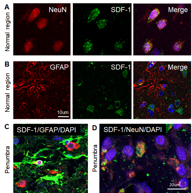
Figure 2.

Phenotypes of SDF-1-positive cells in the human ischemic brain. A-B) Confocal image of representative immunofluorescent staining for NeuN (A) or GFAP (B) (Alexa Fluor 594, red), SDF-1 (Alexa Fluor 488, green), nuclei (DAPI, blue), and merged image from adjacent normal regions of human ischemic stroke brain. C) Merged confocal images of double-label immunohistochemistry in the peri-infarct region (penumbra) of the human ischemic brain section using anti-GFAP (green) and anti-SDF-1 (red). D) Merged confocal images of double-label immunohistochemistry in the penumbra on the human ischemic brain using anti-NeuN (red) and anti-SDF-1 (green). DAPI (blue) was used for nuclei counterstains.
