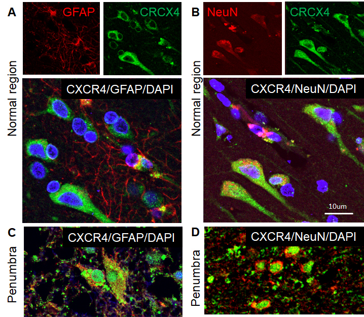
Figure 4.

Phenotypes of CXCR4-expressed cells in the human ischemic brain. Double immunocytochemistry was performed on the ischemic brain sections and the images were recorded using a 2-photon confocal microscope. Representative images show that CXCR4 (green) was expressed in GFAP-positive astrocytes (red) in the normal region (A) and penumbra (C), and NeuN-positive neuronal cells (red) in the normal region (B) and penumbra (D) of human ischemic brain. DAPI (blue) was used for nuclei counterstains.
