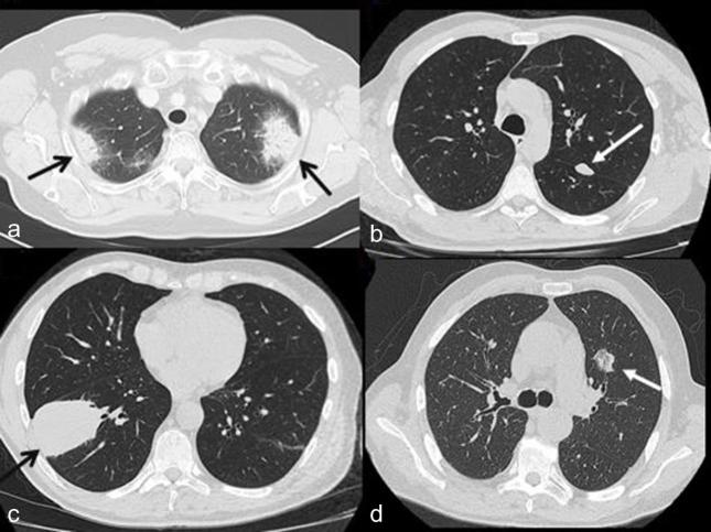
Figure 1.
Example of the main four morphological pattern of presentation of lung lesion at chest CT. (a) Axial CT scan revealing two areas of consolidation in both upper lobes (black arrows) in a 71-year-old female. (b) Axial CT scan showing a nodule of 18 mm of diameter in the upper lobe of left lung (white arrow) in a 58-year-old male. (c) Axial CT scan revealing a big mass of 74 mm of diameter in the lower lobe of right lung (black arrow) in a 58-year-old male (d) Axial CT scan showing a part solid GGO in the left lung (white arrow) in a 72-year-old male.

