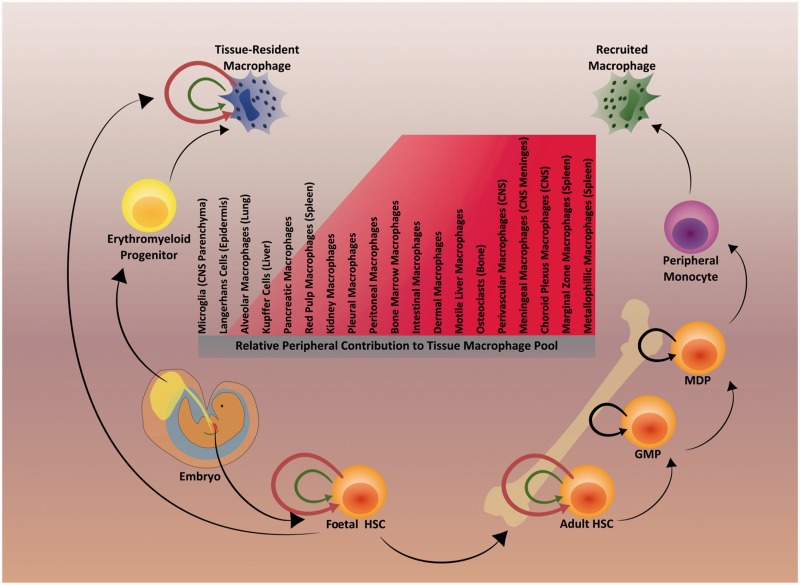
Figure 1.
Primitive and definitive haematopoietic contributions to specific tissue macrophage populations. Some tissue macrophages such as microglia are derived from yolk sac erythromyeloid progenitors during primitive haematopoiesis in early embryonic development. In some cases (e.g. Langerhans cells), foetal progenitors within the liver can also contribute to tissue macrophage populations. Tissue macrophage populations are maintained in the adult organism by local self-renewal and/or monocytes recruited from the periphery. In some cases (e.g. microglia) local self-renewal is the only method by which maintenance occurs. Middle: The contribution of peripheral myeloid cells to the maintenance of specific tissue macrophage populations is tissue-dependent. Here, it is shown as a list with peripheral contribution to specific tissue macrophage populations increasing from left to right. Near the end of the list, populations of tissue macrophages that are completely maintained by peripheral monocytes are shown (e.g. intestinal macrophages and dermal macrophages). Some types, such as white pulp macrophages (spleen), adipose-associated macrophages, and interstitial macrophages (lung) have an undetermined maintenance mechanism and are not depicted (Sieweke and Allen, 2013; Haldar and Murphy, 2014). Pleural macrophages (lung) and peritoneal macrophages each have two distinct subpopulations: a large majority, which originates from the yolk sac is self-maintained, while a smaller group is maintained by monocytes from the periphery (Haldar and Murphy, 2014). The general mechanisms of primitive and definitive haematopoiesis are shown. Tissue-resident macrophages, such as foetal and adult haematopoietic stem cells (HSC), undergo low rates of homeostatic proliferation to self-maintain their populations (represented by green arrows), and may undergo high rates of proliferation when challenged (injury, infection, irradiation, etc. represented by red arrows) (Sieweke and Allen, 2013). Black arrows represent the transient amplification of granulocyte-macrophage (GMP) and macrophage-dendritic progenitors (MDP) during normal haematopoiesis. Overall the majority of these findings come from experiments performed in rodents.

