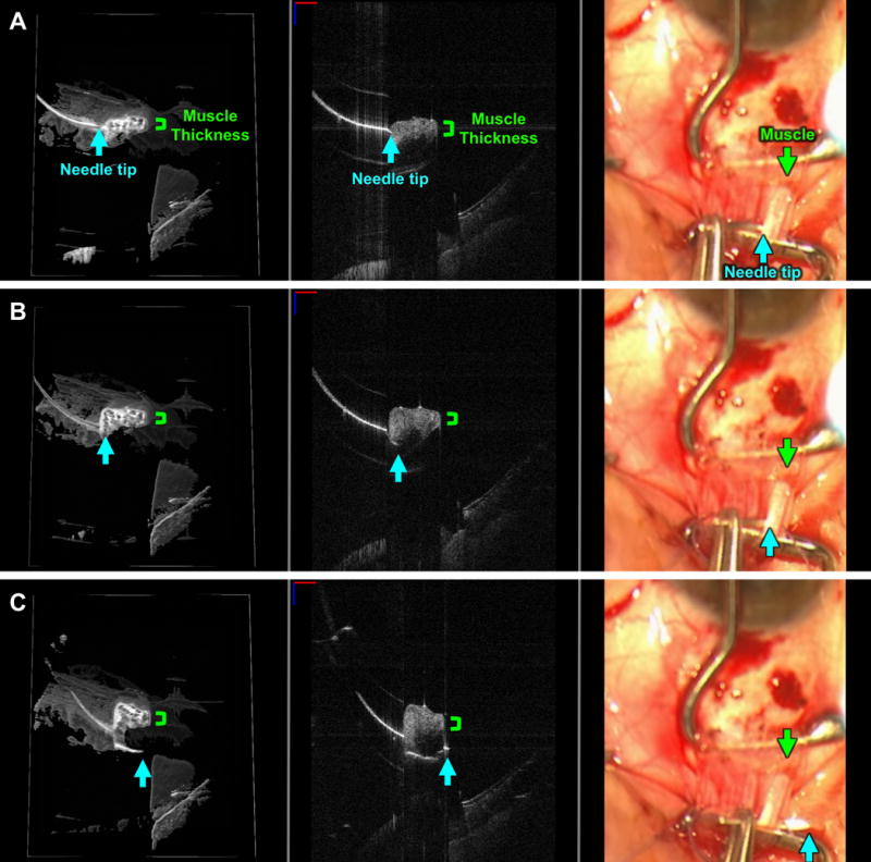
FIGURE 2. Live visualization of a full thickness pass through one pole of the rectus muscle (locking bite) as seen with swept-source microscope-integrated optical coherence tomography (SS-MIOCT).

SS-MIOCT 3D volume (left) with a white box demarcating its corresponding 2D B-scan (middle) and the standard surgical microscope view (right) showing the needle tip (blue arrow) at the surface interface of the lateral rectus muscle (A), progressing beyond the thickness (green bracket) of the muscle (B), and exiting on the other side of muscle (C). The depth of the needle relative to the muscle is much more easily visualized in the 3D volumes and 2D B-scans compared to the standard surgical microscope views (A-C). The suture needle can artifactually appear discontinuous due to different refractive indices of air and tissue. In all SS-MIOCT images, the red scale bar in the 2D B-scan measures 1 mm laterally and the blue scale bar measures 1 mm axially.
