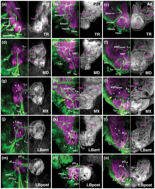
Figure 5.
Metamorphosis of the SEZ. All panels show Z-projections of frontal confocal sections of pupal brain at 12hrs APF (P12; left column; a, d, g, j, m), 24hrs (P24; middle column; b, e, h, k, n) and adult brain (Ad; right column; c, f, i, l, o). Preparations are labeled with anti-Neuroglian (green on left halves of panels; labels secondary axon tracts), and anti-DN-cadherin (neuropil; magenta on left halves of panels; white on right halves). Z-projections represent different levels along the antero-posterior axis [(a–c) tritocerebrum (TR); (d–e) mandibula (MD); (g–i) maxilla (MX); (j–l) anterior labium (LBant); (m–o) posterior labium (LBpost)]. Hatched lines indicate boundaries between columnar neuropil domains, as described in the text. White arrows point at DN-cadherin-rich core of central neuropil column, targeted by lineages 3 and 7 of all neuromeres. White arrowheads point out DN-cadherin-poor poor periphery of central neuropil column, which corresponds to bundles of long axons (CITd and CITv tracts). Blue arrowhead in (g, h) shows chiasmatic midline crossing of lineage 7MX. Large yellow arrow in (h, i) indicates location where DN-cadherin-poor CITd and CITv converge at the later neuropil surface. Small yellow arrows point at DN-cadherin-poor CITv bundle. For abbreviations see List of Abbreviations. Bar: 25μm

