Figure 7.
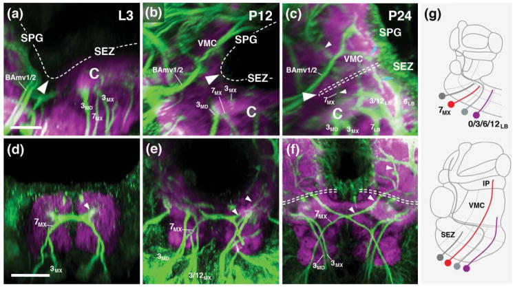
Coalescence of supraesophageal (SPG) and subesophageal neuropils (SEZ). (a–f) Z-projections of parasagittal (a–c) and frontal (d–f) confocal sections of third instar larval brain (a, d), 12hr pupal brain (b, e) and 24hr pupal brain (c, f) labeled with anti-Neuroglian (green; labels secondary axon tracts), and anti-DN-cadherin (neuropil; magenta). Hatched line in (a–c) and (f) demarcates dorsal surface of neuropil. Large white arrowheads point at boundary between SPG and SEZ. Note posterior tilt of SPG between larval and 24hr pupal stage. From 24hr pupal development onward, the dorsal deuterocerebrum (called the ventromedial cerebrum (VMC) in the standard nomenclature for the adult brain), which forms the most ventral part of the SPG, covers the dorsal surface of the SEZ [hatched double line in (c, f)]. Also note growth of tract of lineage 7MX [small white arrowheads in (c–f)]) into SPG. Blue arrows in (c) point at tract of lineage 12LB, which also reaches into the SPG. (g) Schematic sagittal section of late larval brain (top) and pupal brain (bottom), depicting extension of SEZ lineages 7MX and 0/3/6/12LB into SPG. For other abbrveviations, see List of Abbreviations. Bar: 10μm (a–c); 25μm (d–f).

