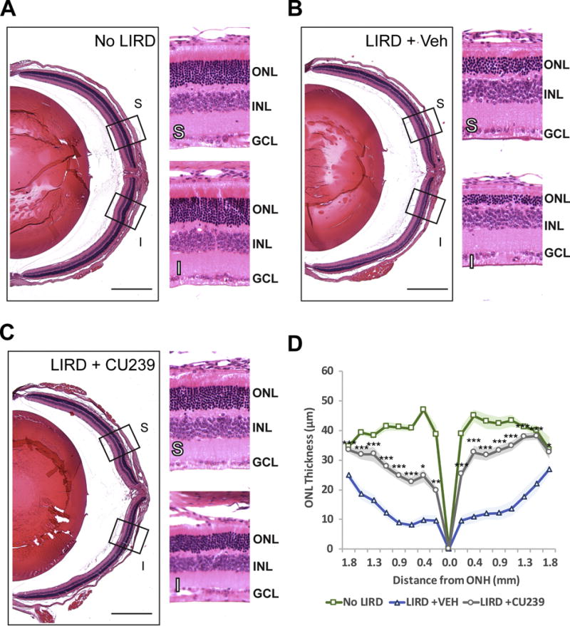
Fig. 6. Histological analysis of the retinas from CU239 injected mice 5 days post-LIRD.

Retinal cross-sections of (A) No LIRD Control, (B) LIRD + Vehicle, and (C) LIRD + CU239 were stained with H&E for morphological comparison. (D) A graph showing ONL thickness measured in cross sections of the retina. Student’s t-test. *P<0.05 and ***<0.001. (Mean ± SEM). S: Superior retina; I: inferior retina. Scale bar: 500 μm (4× images). Boxed areas on 4× images were zoomed under 20× magnification.
