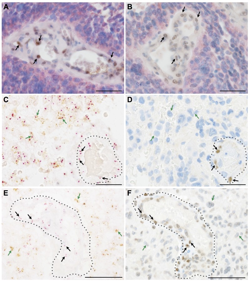
Figure 3. Key EMT molecules are expressed by non-neoplastic vessel-associated cells in human astrocytomas.
(A, B) Double immunohistochemistry for mutated IDH-1 (R132H) (red) as a marker for neoplastic glial cells in a secondary glioblastoma together with (A) SLUG (brown, arrows) or (B) TWIST (brown, arrows) indicating that EMT molecules are restricted to IDH-1 (R132H)-negative, vessel-associated cells. (C, E) Silver in-situ hybridization (SISH) staining (brown dots; green arrows) for EGFR-amplification indicating glial cells in primary glioblastoma and chromosome 7 control probes (red dots; red arrows) combined with (D) SLUG and (F) TWIST immunohistochemistry showing that mutated glioma cells are negative for EMT molecules, the latter being present on tumor-associated blood vessels (dotted lines; scale bars: A-F 50μm).

