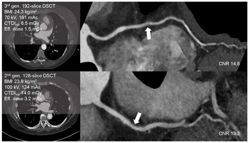
Fig 4.
Example clinical cases of quantitative contrast-to-noise ratio measurements demonstrating representative image quality on the 3rd generation DSCT (above) and 2nd generation DSCT (below) on patients with similar BMI. Curved planar reformat images demonstrate noncalcified plaque resulting in mild stenosis of the right coronary artery (right, arrows).

