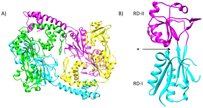
Figure 1. Crystal Structure of AphB.
A) Ribbon structure of the AphB tetramer. Extended subunits are shown in cyan and magenta and compact subunits are shown in green and yellow (PDB ID: 3SZP). B) Monomer of the regulatory domain of AphB. The previously identified putative effector-binding pocket (denoted with an asterisk) is located between RD-I (cyan) and RD-II (magenta).16

