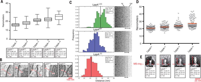
Fig. 3. The inner-to outer-membrane distance and flagellar rod length varied with LppA lengths.

Resin-embedded LppA-length mutants, as well as WT control (TH22579 and TH22634 to TH22637) were thin-sectioned and observed by electron tomography. (A and B) The peptidoglycan-layer (PG)–to–outer-membrane (OM) distances for each strain were measured. As the length of LppA increased, we observed a corresponding increase in PG-to-OM distance [P < 0.0001, one-way analysis of variance (ANOVA)]. (C) To verify these results, cells from −21, +21, and WT LppA strains (TH22579, TH22634, and TH22635) were imaged via cryo-EM, and the distances between the inner membrane (IM) and OM were measured for each. The distances varied with LppA lengths (P < 0.0001, Student’s two-tailed t test). (D and E) Flagellar rods from lengthened LppA variants (TH22574 to TH22577) were purified, imaged via TEM and measured. The average length of the rod increased ~1.5 to 2 nm for every three heptad repeats (21 residues) added [P < 0.0001, one-way ANOVA, N = ≥ 3, data in (D) are means ± SEM].
