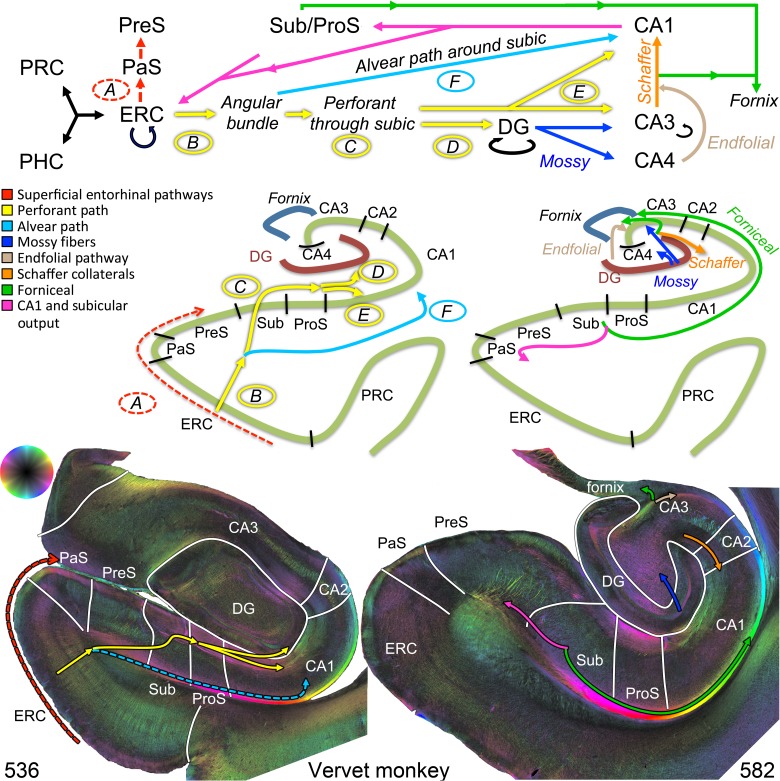
Figure 1.
Hippocampal circuitry and validation in the vervet monkey. Top: A–F indicate the components of the perforant path system largely identified from animal studies. Major pathways are represented by solid lines, whereas minor pathways are represented by dotted lines. A = “sublamina supratangentialis” (dotted red), a minor superficial entorhinal bundle projecting to the parasubiculum and presubiculum. B = entorhinal projection to the angular bundle (yellow). C = angular bundle fibers (yellow) perforating through the subiculum. D + E = these same fibers projecting superiorly (D) into the dentate gyrus or inferiorly (E) into the hippocampal CA fields (yellow). F = angular bundle fibers subjacent to the subiculum projecting into the CA fields (alvear path, light blue: note, for the human diagram, this pathway is larger in humans and is depicted as solid, whereas it is smaller in monkeys and depicted as dotted light blue on the bottom left). The remaining hippocampal circuitry includes: mossy fibers projecting from the dentate gyrus to CA3 and CA4 (dark blue), the endfolial pathway projecting from CA4 toward CA3 (light brown), CA3 projecting to the fornix (green) and to CA1 (Schaffer collaterals, orange), followed by CA1 and subiculum output to ERC (pink), and subicular output to the fornix (green). CA, cornu ammonis; DG, dentate gyrus; ERC, entorhinal cortex; PaS, parasubiculum; PHC, parahippocampal cortex; PRC, perirhinal cortex; PreS, presubiculum; ProS, prosubiculum; Sub, subiculum. Bottom: Polarized light microscopic images from the vervet monkey illustrate the above pathways.

