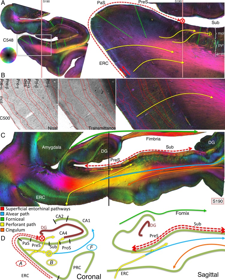
Figure 3.
Entorhinal pathways and the angular bundle. (A) Coronal PLI at the level of the posterior hippocampal head of the left hemisphere of brain #1. The approximate localization of sagittal section S190 (shown in C) is indicated by peach vertical line. The green boundaries correspond to the subregion demarcations on the transmittance images from Figure 2E. The hue on the color wheel indicates the direction of the in-plane fiber orientation (see Supplementary Fig. 1), and the brightness/darkness of the color (e.g., more peripheral/central in the color wheel) indicates a primarily in-plane/through-plane orientation. Arrows with cross hairs denote a through-plane orientation. Best seen on the zoomed in coronal section from the box on the left are dual superficial entorhinal pathways (red arrows) running tangentially throughout the ERC to the parasubiculum and presubiculum. Both project longitudinally in the molecular layer of the presubiculum (red cross hair), confirmed on sagittal plane S190 of the right hemisphere in (C). In yellow are medial entorhinal projections to the angular bundle (lower 2 curvilinear yellow arrows) and from the angular bundle through the presubiculum into the molecular subiculum (top yellow sigmoidal arrows). Subicular lamination on the far right is indicated as mol, molecular; pyr, pyramidal; a–m, alvear/multiforme. (B) A slightly more anterior section (C500) with Nissl staining performed after PLI for visualization of cell bodies. Lamination of the ERC derived from the Nissl stain is superimposed on the transmittance and fiber orientation maps. Perforant pathway fibers originate in approximately layer Pre-α of the ERC and radially project to the angular bundle (yellow curved arrow). (C) Sagittal PLI slice S190 from the right hemisphere of brain #1 at the approximate location indicated by the vertical peach line in A. The approximate localization of the coronal section from the left hemisphere in A is indicated by the vertical black line on this right sagittal section. Red arrows highlight the presubicular longitudinal components of the superficial entorhinal pathways from A. The lower yellow arrow signifies perforant path projections from the medial ERC, through the angular bundle, and toward the presubiculum and subiculum. More superiorly is a longitudinal entorhinal pathway (thin yellow arrow) running in the pyramidal layer of the presubiculum, depicted as the yellow ellipses in Figure 4. The green arrow depicts fibers projecting to the fimbria; in blue is the alvear bundle to the posterior hippocampus; and in orange is the cingulum bundle within the parahippocampal gyrus (see Supplementary Fig. 3). (D) Diagrammatic representation of entorhinal pathways in the coronal and sagittal planes.

