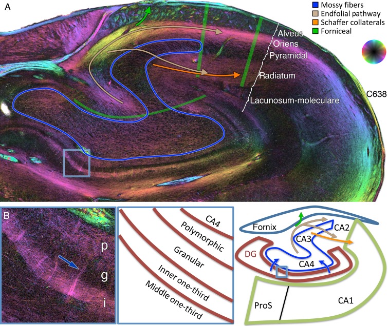
Figure 6.
Dentate fibers, mossy fibers, endfolial pathway, and Schaffer collaterals. (A) Coronal PLI at the level of the hippocampal body showing a relatively diminished color orientation in fields CA4 and CA3 pyramidal cell layers, circumscribed in blue, representing the unmyelinated target of the mossy fibers. Endfolial fibers originate in CA4 (upper light brown curvilinear arrow) and project to stratum oriens. The Schaffer collaterals originate in CA3 (orange arrow) and project to the stratum radiatum. Fibers project from the endfolial pathway to the Schaffer collaterals (lower light brown sigmoidal arrow). The overlying alveus contains largely longitudinal forniceal fibers (see Figs 3C and 5A,B). (B) Zoomed in section on the medial and inferior dentate gyrus indicated by the blue box in A at the bottom left. p, polymorphic layer, g, granular cell layer, and i, inner one-third of the dentate gyrus. This demonstrates tangential fibers in the polymorphic layer and inner one-third of the dentate gyrus, likely representing mossy cell associational fibers. Some subtle fibers can be seen traversing the granular layer to bridge these 2 layers (dark blue arrow).

